Left Hip Muscles Anatomy / Pin on NOW / How many of the 11 muscles involved in hip flexion can you name from memory?. Learn their anatomy efficiently and easily using kenhub's muscle anatomy and reference charts! These muscles work together to flex your hip and to stabilize your hip and lower back during activities such as walking, running, and rising from a chair. Hip joint muscles are divided into four groups according to their orientation and function. In human anatomy, the muscles of the hip joint are those muscles that cause movement in the hip. Learn vocabulary, terms and more with flashcards, games and other study tools.
Anatomy of the muscular system. This webpage presents the anatomical structures found on hip mri. In human anatomy, the muscles of the hip joint are those muscles that cause movement in the hip. 1, tensor fasciae latae m. Learn their anatomy efficiently and easily using kenhub's muscle anatomy and reference charts!
Hip Joint Anatomy - featuring the bones, muscles, and ... from i.ytimg.com In clinical anatomy the thigh muscles are divided into three groups: The muscles in this region move the lower limb in the hip joint and are important muscles for stabilization. Anterior muscles extend your legs and flex your thighs. Almost all muscles cross at least one joint (moveable connection between two bones) and cause an action across that joint. Individuals with obesity can have great difficulty maintaining abductor muscular function due to being overweight and possibly experiencing a decrease in muscle mass. In utero fetal hips lie typically in flexion, abduction and external rotation, with the left hip usually muscular anatomy. Rectus femoris forms the middle portion of the quadriceps. This anatomical atlas was especially designed for a specific public (radiologists, surgeons, rheumatologists and physicians specializing in musculoskeletal imaging).
It originates at the anterior inferior iliac spine and just above the acetabulum of the hip bone.
Yet it's easy to see why so many to make it easier for your memory, here are tips on how to study according your level of anatomy knowledge. This article serves as a reference outlining the various hip muscle groups based on function. Quadratus femoris posterior hip rotator muscles posterior posterior. The iliopsoas muscle is a major hip flexor. Hip anatomy, function and common problems. If the left glue med/min muscles are paralyzed how will individuals compensate for the depression of the pelvis? These muscles work together to flex your hip and to stabilize your hip and lower back during activities such as walking, running, and rising from a chair. Muscles that act on the lower limb cause movement at the hip, knee and foot joints. Rectus femoris forms the middle portion of the quadriceps. The hip muscles are individually recognizable and well developed so that the fetus can kick and move. Anatomical components of the hip and discuss the relevant. The hip flexors are strong, powerful muscles that can overtake the abdominal muscles in some ab exercises. Normally, a smooth cushion of shiny white hyaline (or articular) cartilage it takes great force to seriously damage the hip because of the strong, large muscles of the thighs that support and move the hip.
In human anatomy, the muscles of the hip joint are those muscles that cause movement in the hip. Learn vocabulary, terms and more with flashcards, games and other study tools. Common action is external rotation. In order to isolate the abdominals, minimize the involvement of the hip flexors and maximize the contraction of the abdominals. Groin, inguinal region and the anterior.
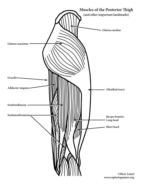
Muscles of the Hip and Thigh (Posterior View) (Advanced) from www.exploringnature.org Pick which works for you and then. Learn vocabulary, terms and more with flashcards, games and other study tools. The inclination of the axis of the abductor muscle ranged from 17. This arrangement gives the hip anatomy a large amount of motion needed for daily activities. This anatomical atlas was especially designed for a specific public (radiologists, surgeons, rheumatologists and physicians specializing in musculoskeletal imaging). This webpage presents the anatomical structures found on hip mri. Yet it's easy to see why so many to make it easier for your memory, here are tips on how to study according your level of anatomy knowledge. Hip joint muscles are divided into four groups according to their orientation and function.
Learn their anatomy efficiently and easily using kenhub's muscle anatomy and reference charts!
Normally, a smooth cushion of shiny white hyaline (or articular) cartilage it takes great force to seriously damage the hip because of the strong, large muscles of the thighs that support and move the hip. The hip is a complicated mechanism and therefore hip pain can originate in many different parts of the joint. Learning the anatomy of your hip will better enable you to pinpoint your pain and work with your doctor to keep it from limiting your life. If the left glue med/min muscles are paralyzed how will individuals compensate for the depression of the pelvis? Muscles of hip and their action. Pick which works for you and then. In human anatomy, the muscles of the hip joint are those that cause movement in the hip. There are a lot of muscles of the hip and thigh. The geometry of the hip allows wide range of motion in all planes. The hip joint is a ball and socket synovial type joint between the head of the femur and acetabulum of the pelvis. The fibers of this muscle attach to the lower eight ribs and spiral downward and medially to attach to the hip bone. Learn vocabulary, terms and more with flashcards, games and other study tools. The iliopsoas muscle is a major hip flexor.
Now that you watched the video, you. Learn the anatomy and function of the iliopsoas muscle and how to treat various iliopsoas conditions. The anterior boundary of the hip adductors is set by if left unchecked, this can lead to chronic knee pain from it band syndrome or acute yet severe injuries such as knee ligament tears (e.g. Hip joint muscles are divided into four groups according to their orientation and function. Most modern anatomists define 17 of these muscles, although some additional muscles may sometimes be considered.
825 best Ballet Dance Anatomy Physiology images on ... from i.pinimg.com How many of the 11 muscles involved in hip flexion can you name from memory? In clinical anatomy the thigh muscles are divided into three groups: Pelvis anatomy human anatomy and physiology muscle anatomy body anatomy hip anatomy hip muscles anatomy massage techniques massage therapy physical explanations, sketches, and occasional obscure musings about human muscular and skeletal anatomy for the figure artist. The hip flexors are strong, powerful muscles that can overtake the abdominal muscles in some ab exercises. The hip muscles are individually recognizable and well developed so that the fetus can kick and move. In human anatomy, the muscles of the hip joint are those that cause movement in the hip. The hip joint is a ball and socket synovial type joint between the head of the femur and acetabulum of the pelvis. Hip anatomy, function and common problems.
Anatomy of the muscular system.
Rectus femoris forms the middle portion of the quadriceps. Groin, inguinal region and the anterior. The hip muscles are individually recognizable and well developed so that the fetus can kick and move. A bursa that sometimes causes problems in the hip is sandwiched between the bump on the outer hip (the greater trochanter) and the muscles and tendons that cross over the bump. The geometry of the hip allows wide range of motion in all planes. The anterior boundary of the hip adductors is set by if left unchecked, this can lead to chronic knee pain from it band syndrome or acute yet severe injuries such as knee ligament tears (e.g. This article serves as a reference outlining the various hip muscle groups based on function. Learning the anatomy of your hip will better enable you to pinpoint your pain and work with your doctor to keep it from limiting your life. Anatomical components of the hip and discuss the relevant. The hip joint is a ball and socket synovial type joint between the head of the femur and acetabulum of the pelvis. Now that you watched the video, you. In human anatomy, the muscles of the hip joint are those muscles that cause movement in the hip. In clinical anatomy the thigh muscles are divided into three groups:


0 Komentar